baby chest x ray radiation
Lung tissue absorbs little radiation and will appear dark on the image. Fetal doses resulting from radiological examination of the mothers skull head neck chest and extremities are extremely low 001 rad because of the relatively low maternal radiation dose beam direction and distance between the primary field and the fetus.
Radiation From X Rays And Living In Denver They Re Not The Same
However radiation therapy has been shown to decrease milk production in some women and.

. All distal extremity exposures are taken at 110115 cm SID. From an x-ray is of clear benefit to the mother and baby the examination will be done with a minimal dose to the fetus well below the level of injury to the fetus. X-rays are a form of radiation.
The examination should only proceed after approval by a Radiologist. Long-term problems are very small. In general the amount of radiation used for simple chest x-rays is so small that its considered safe during pregnancy but your healthcare specialist can help you make the decision to perform the x-ray based on the urgency of your symptoms.
CT scans can deliver radiation doses that are up to 200 times higher than an average chest X-ray. X-rays are used to make pictures of the bones and organs. CT Scan of your chest with lead apron over abdomen Between 3 and 6 tests.
Type of X-ray or scan Quantity X-ray of your pelvis Between 5 and 30 tests. Radiation exposure from X-rays does not pose any short-term problems. One single chest x-ray is not concerning at all--even to newborns.
Most researchers agree that babies who receive a small dose of radiation equal to 500 chest x-rays or less at any time during pregnancy do not have an increased risk for birth defects. 43 rows Like other sources of background radiation the amount of radon exposure varies widely depending. If the mother is undergoing low dose radiation therapy on a localized area according to the British Journal of Radiology the mother can still choose to breastfeed if both the mother and babys doctor feel it is safe and no potential risk is posed to the infants over all health.
Most x-ray images are electronically stored digital files. At Stanford we take extra precautions to minimize our patients exposure to radiation including using. On a chest x-ray the ribs and spine will absorb much of the radiation and appear white or light gray on the image.
Radiation exposure can cause harm to a growing babys developing baby. X-ray of your lower spine Between 3 and 20 tests. When the X-rays pass through the body they create an image like a shadow.
Ionising radiation and X-rays Ionising radiation which can be either particle or electromagneticconsists of individual particles. The only increased risk to these babies is a slightly higher chance of having cancer later in life less than 2 higher than the normal expected. Erect chest X-rays are taken at 180 cm.
Different parts of the body contain different tissues which vary in how much X-ray radiation they absorb depending on how dense they are. Radiation exposure is known to damage the cells that were exposed and can lead to cancer. The radiation exposure of an adjacent newborn the radiographer and other persons in the room was simulated using phantoms during X-ray examination of the chest using vertical and horizontal beams.
This kind of radiation is invisible. Food and Drug Administration. Full legfull spine imaging is performed at 180 cm using CR.
VQ Lung Scan Approximately 5 or more tests CT Scan of your head with lead apron over abdomen Approximately 50 tests. Not providing the proper protection during the X-ray or over-radiating the baby can cause serious harm. By definition exposure is a measure of the amount of ionizations produced in air by photon radiation.
It is not putting your child at any risk. Approximately 1 in 500 children develops cancer during childhood even without radiation as a fetus. CT Scan of your abdomen.
The Alliance for Radiation Safety in Pediatric Imaging reminds parents and pediatricians to follow these guidelines. And thats not counting the very common follow-up CT scans. Your doctor can easily access these stored images to diagnose and manage your condition.
Free Healthy Baby App for iPhone. Most of the increased exposure in the United States is due to CT scanning and nuclear imaging which require larger radiation doses than traditional x-rays. To evaluate the degree of radiation exposure from diagnostic X-ray examinations with mobile X-ray machines in a premature intensive care unit.
X-Rays Scans Radiation and Kids. Eg if you have a chest x ray you are exposed to radiation. Radiation exposure from X-rays may slightly raise the risk of later cancer especially in children who have had many tests with high radiation exposure.
Exposure is commonly used to refer to being around a radiation source. Lateral cervical spines are taken at 150 cm. Neonates in a special care baby unit SCBU often require frequent chest and abdomen radiographs in a short period of time to monitor the treatment progress of the neonate and to check the postion of the various tubes and catheters used in SCBU Spiegel 1995.
They have been associated with a very small increased risk of cancer especially leukemia for an unborn baby especially when done later in pregnancy in large amounts. But the risk is very small. Risks depend on the amount of radiation to which the baby was exposed and the amount of time that it was exposed.
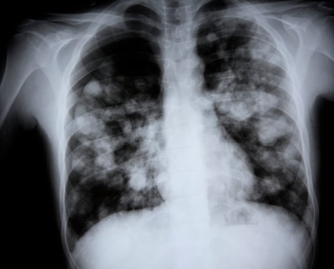
A chest X-ray is a painless noninvasive procedure with few risks. An X-ray is a picture which is taken using a form of radiation that is able to pass through the body to create a digital X-ray image. X-rays use a small amount of radiation about the same levels that occur naturally in the environment.
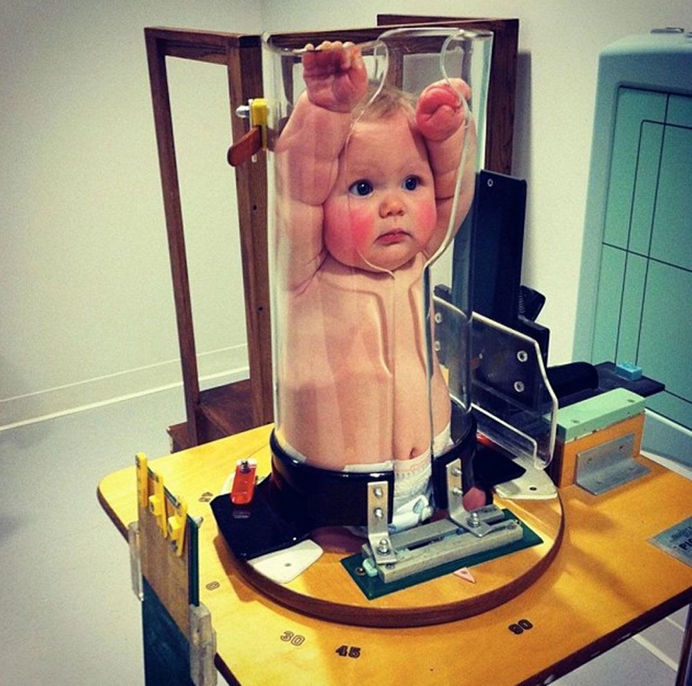
A protective lead apron to shield certain parts of the body. For example if the radiation dose to the unborn baby was roughly equivalent to 500 chest x-rays at one time the increase in lifetime cancer risk would be less than 2 above the normal lifetime cancer risk of 40 to 50. Burns hair loss and an increased risk of cataracts are also symptoms of excess radiation exposure according to the US.
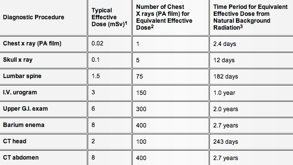
Imaging should only be used if there is a definite medical benefit. Radiographic exposure of neonates attracts particular interest because of their greater cell. A chest x-ray for example delivers 01 mSv while a chest CT delivers 7 mSv see the table 70 times as much.
1 A recent study claiming an association between dental radiography in pregnancy and low birth weight. Including conventional X-rayintravenous pyelographyventilationperfusion V-P lung scanningangiography and computed tomography CTand the safety of contrast media in diagnostic procedures in pregnancy.

What Is An X Ray For Kids Radiology And Medical Imaging

Is A Single Chest X Ray Too Much Radiation For A 3 Year Old Healthychildren Org

Diagnostic Imaging
Simple Diagnostic Model For Pneumonia In Kids To Reduce Need For X Rays Imaging Technology News

Pem Pearls Chest Radiographs For Shortness Of Breath

Neonatal Chest Radiography Influence Of Standard Clinical Protocols And Radiographic Equipment On Pathology Visibility And Radiation Dose Using A Neonatal Chest Phantom Radiography

Radiation Doses In Neonatal X Ray Examinations Download Table

Children S National Nicu Reduces Chest X Rays Unintended Extubations Innovation District Children S National

X Rays And Unshielded Infants Raise Alarms The New York Times

A Pediatric Chest X Ray With The Pb Shield The Circles Are The Download Scientific Diagram

X Rays And Unshielded Infants Raise Alarms The New York Times

Pediatric Chest Supine View Radiology Reference Article Radiopaedia Org
This Adorable Baby Is Squished Into A Tube For A Good Reason

Indications For Chest X Rays In Children And How To Obtain And Interpret Them

Paediatric Chest Pa Erect View Radiology Reference Article Radiopaedia Org
Lifetime Cancer Risks From X Rays For Children Relatively Low

Chest X Rays For Children Doctors Are Unnecessarily Exposing Kids To Radiation With No Clinical Benefit

X Ray For Kids Children S Health Orange County

Pdf Radiation Protection In Pediatric Radiography Introducing Some Immobilization And Protection Equipment Semantic Scholar